وبلاگ جامع علوم پرتو پزشکی و فیزیک پزشکی

پرتو پزشکی, فیزیک پزشکی, ام آر آی MRI ,سی تی اسکن Ct Scan , رادیولوژی, رادیوتراپی, پزشکی هسته ای, مهندسی هسته ای

وبلاگ جامع علوم پرتو پزشکی و فیزیک پزشکی

پرتو پزشکی, فیزیک پزشکی, ام آر آی MRI ,سی تی اسکن Ct Scan , رادیولوژی, رادیوتراپی, پزشکی هسته ای, مهندسی هسته ای

۹ مطلب با موضوع «Salient Points» ثبت شده است

دز تابش در یک ماموگرافی غربالگری روتین در خانم ها چه مقدار می باشد؟

مقدار تابش برابر با 0.2 Rad و یا 2 mGy برای دونما  از یک Breast می باشد.

هر چند در این روش بیمار در برابر تابش قرار می گیرد اما مقدار این تابش به طور قابل ملاحظه ای کم بوده و اعتقاد بر این است که مزایای تشخیص زود هنگام بر معایب آن بیشتر می باشد.


رفرنس:

Radiology secrets plus 4th ed
 

  • medradiation

Relative Biological Effectiveness

medradiation | شنبه, ۲۹ مهر ۱۳۹۶، ۰۷:۲۱ ق.ظ

Relative Biological Effectiveness (RBE)

The relative biological effectiveness (RBE) of a radiation under test (e.g. a high-LET radiation) is defined as:


to give the same biological effect. The reference low-LET radiation is commonly 250 kVp X-rays or Co60γ-rays since these radiations are usually available whenever RBE is being evaluated.


Basic Clinical Radiobiology

Download


  • medradiation

CEMA

medradiation | دوشنبه, ۳۰ مرداد ۱۳۹۶، ۰۶:۴۶ ق.ظ

CEMA

Cema is the acronym for converted energy per unit mass. It is a nonstochastic quantity applicable to directly ionizing radiations such as electrons and protons. The cema C is the quotient of dEc by dm, where dEc is the energy lost by charged particles, except secondary electrons, in collisions in a mass dof a material:

C = dEc / dm


The unit of cema is joule per kilogram (J/kg). The name for the unit of cema is
the gray (Gy).


Radiation Oncology Physics A Handbook for Teachers And Students
 

  • medradiation

KERMA

medradiation | چهارشنبه, ۲۷ بهمن ۱۳۹۵، ۰۴:۱۱ ب.ظ
KERMA

Kerma is an acronym for kinetic energy released per unit mass. It is a nonstochastic quantity applicable to indirectly ionizing radiations such as photons and neutrons. It quantifies the average amount of energy transferred from indirectly ionizing radiation to directly ionizing radiation without concern as to what happens after this transfer.
  • medradiation

Scatter Air Ratio

medradiation | پنجشنبه, ۲۳ ارديبهشت ۱۳۹۵، ۱۰:۴۶ ق.ظ

SARs are used for the purpose of calculating scattered dose in the medium. The computation of the primary and the scattered dose separately is particularly useful in the dosimetry of irregular fields.

SAR may be defied as the ratio of the scattered dose at a given point in the phantom to the dose in free space at the same point. The SAR, like the TAR, is independent of the SSD but depends on the beam energy, depth, and field size.
Because the scattered dose at a point in the phantom is equal to the total dose minus the primary dose at that point, SAR is mathematically given by the difference between the TAR for the given field and the TAR for the 0 × 0 field

SAR (d, rd) = TAR (d, rd) - TAR (d,0)

Here TAR (d,0) represents the primary component of the beam.


Khan's The Physics of Radiation Therapy Fifth Edition

Download

  • medradiation

TAR or Tissue Air Ratio

medradiation | پنجشنبه, ۱۹ شهریور ۱۳۹۴، ۰۹:۲۴ ق.ظ

Tissue-Air Ratio


Tissue-air ratio (TAR) was first introduced by Johns  in 1953 and was originally called the “tumor-air ratio.” At that time, this quantity was intended specifically for rotation therapy calculations. In rotation therapy, the radiation source moves in a circle around the axis of rotation, which is usually placed in the tumor. Although the SSD may vary depending on the shape of the surface contour, the source-axis distance remains constant.
Since the percent depth dose depends on the SSD , the SSD correction to the percent depth dose will have to be applied to correct for the varying SSD—a procedure that becomes cumbersome to apply routinely in clinical practice. A simpler quantity—namely TAR—has been defined to remove the SSD dependence. Since the time of its introduction, the concept of TAR has been refined to facilitate calculations not only for rotation therapy, but also for stationary isocentric techniques as well as irregular fields.


Tissue-air ratio may be defined as the ratio of the dose (Dd) at a given point in the phantom to the dose in free space (Dfs) at the same point.


This is illustrated in Figure 2 For a given quality beam, TAR depends on depth d and field size rd at that depth:

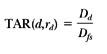
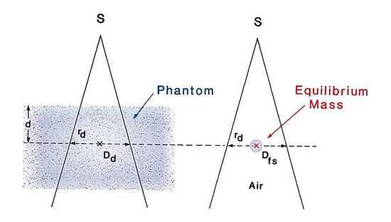
Fig 2: Illustration of the definition of tissue-air ratio (TAR). TAR(d,rd) = Dd/Dfs


Physics of Radiation Therapy, The, 5th Edition

Faiz M. Khan PhD

Professor Emeritus

download


  • medradiation

Percentage Depth Dose

medradiation | يكشنبه, ۸ شهریور ۱۳۹۴، ۱۰:۵۵ ق.ظ
Percentage Depth Dose

One way of characterizing the central axis dose distribution is to normalize dose at depth with respect to dose at a reference depth. The quantity percentage (or simply percent) depth dose may be defined as the quotient, expressed as a percentage, of the absorbed dose at any depth d to the absorbed dose at a fixed reference depth d0, along the central axis of the beam (Fig. 1). Percentage


Figure 1: Percentage depth dose is (Dd/Dd0), where d is any depth and d0 is reference depth of maximum dose.


For orthovoltage (up to about 400 kVp) and lower-energy x-rays, the reference depth is usually the surface (d0 = 0). For higher energies, the reference depth is taken at the position of the peak absorbed dose (d0 =dm).

In clinical practice, the peak absorbed dose on the central axis is sometimes called the maximum dose, the dose maximum, the given dose, or simply the Dmax. Thus:

A number of parameters affect the central axis depth dose distribution. These include beam quality or energy, depth, field size and shape, source to surface distance, and beam collimation.


Physics of Radiation Therapy, The, 5th Edition

Faiz M. Khan PhD

Professor Emeritus

دانلود


  • medradiation

Top Secrets

medradiation | شنبه, ۲۴ مرداد ۱۳۹۴، ۰۸:۵۴ ق.ظ
Top Secrets

  1. PACS stands for picture archiving and communication systems. These are the systems used by digital radiology departments to store, network, and view imaging studies.
  2.  RIS stands for radiology information system. RIS manages patient scheduling and tracking, examination billing, and receipt/display of radiology reports.
  3. Almost all hepatic cysts and hemangiomas can be differentiated from malignant liver disease by the use of heavily T2-weighted (>180 ms) MR images.
  4. On postcontrast images, the normal spleen displays alternating bands of high and low attenuation (CT) or signal (MRI) in the arterial phase. The spleen appears more homogeneous in a more delayed phase.
  5.  Splenic laceration can be differentiated from developmental splenic cleft. Patients with laceration have a trauma history, display a low attenuation defect with sharp edges, and have perisplenic hemoperitoneum.
  6.  MRI and CT are less specific in the characterization of splenic lesions than they are in characterization of liver, adrenal, or renal lesions.
  7.  The pancreatic neck and body are the most common portions of the pancreas to be injured in blunt trauma because they are compressed against the spine in blunt traumatic injuries to the abdomen.
  8.  The most specific CT imaging finding of acute appendicitis is an abnormal appendix that is typically dilated 6 mm or greater and fluid-filled. A calcified appendicolith with periappendiceal fat stranding is another highly specific CT finding.


Radioology secrets plus
برای دانلود کتاب می توانید از لینک زیر استفاده کنید

medradiation.sellfile.ir
  • medradiation

Top Secrets

medradiation | دوشنبه, ۲۹ تیر ۱۳۹۴، ۰۷:۴۵ ب.ظ

Top Secrets

نکات مهم در رادیولوژی - بر گرفته از کتاب اسرار رادیولوژی


  1. Increasing voltage (kV) decreases contrast and increases exposure, making the film darker. Increasing milliampereseconds (mAs) increases exposure, making the film darker.
  2. A scout film should always be obtained before performing a fluoroscopy study with a contrast agent. The scout film
    allows the radiologist to determine whether an object that appears “white” on a radiograph is bone or metal versus
    contrast (the latter would not be on the scout film).
  3. Structures in the body that are very dense (such as structures that contain calcium) attenuate a large amount of the x-ray beam; the x-ray beam is unable to reach the film and darken it, and such structures appear white on a radiograph.Conversely, structures that are not very dense (such as air) allow the x-ray beam to penetrate and darken the film; such structures appear black.
  4. Regions with many acoustic interfaces reflect a lot of sound back to the transducer. These are termed echogenic or
    hyperechoic, and by convention are viewed as bright areas on ultrasound (US). Regions with few acoustic interfaces do not reflect many sound waves; they are termed hypoechoic and are viewed as dark areas.
  5. Electron-dense structures, such as metal and bone, stop a large number of x-rays and are bright on computed
    tomography (CT). Regions with lower electron density, such as air or fat, stop very few x-rays and are rendered as dark.Because CT images are created with x-rays, the same things that are bright and dark on plain films are bright and dark on CT.
  6. T1-weighted images have a short “time to repetition” (TR) (<1000 ms) and a short “time to echo” (TE) (<20 ms).T2-weighted images have a long TR (>2000 ms) and a long TE (>40 ms).
  7. To differentiate between T1-weighted and T2-weighted images, look for simple fluid. Fluid tends to be hyperintense to virtually everything else on T2-weighted images. On T1-weighted images, fluid is of low intermediate signal. Good places to look for fluid include the urinary bladder and the cerebrospinal fluid.
  8. Nuclear medicine is unique in that its strength lies in portraying the functional status of an organ, rather than producing images that are predominantly anatomic in content
  9. In nuclear medicine studies, the radiologist administers a radioactive atom, either alone or coupled to a molecule, that is known to target a certain organ or organs. Its distribution is examined to determine any pathologic condition in that particular organ.

Radioology secrets plus
برای دانلود کتاب می توانید از لینک زیر استفاده کنید
http://medradiation.sellfile.ir
  • medradiation